


AI Breakthrough: Early Osteoporosis Detection Revolutionizes Healthcare
Researchers at geneonline.com have developed an artificial intelligence technique leveraging knowledge distillation to improve the accuracy of detecting low bone mass, a key indicator of osteoporosis, from chest X-rays. The advancement, announced on October 26, 2023, could lead to earlier diagnosis and intervention for millions at risk.
Background: The Challenge of Early Detection
Osteoporosis, a condition characterized by weakened bones, affects an estimated 46 million Americans, with a significant portion remaining undiagnosed until a fracture occurs. Traditional methods for assessing bone density, such as dual-energy X-ray absorptiometry (DEXA) scans, are often expensive and not readily accessible to everyone. Chest X-rays are routinely performed for various respiratory conditions, presenting an opportunity to potentially screen for early signs of bone loss. However, accurately identifying subtle changes in bone density from these images has been a persistent challenge for radiologists.
For years, the field has explored using machine learning to assist in medical image analysis. Early attempts relied on training models directly on labeled chest X-ray datasets showing bone density variations. However, these models often struggled with limited data availability and variations in image quality.
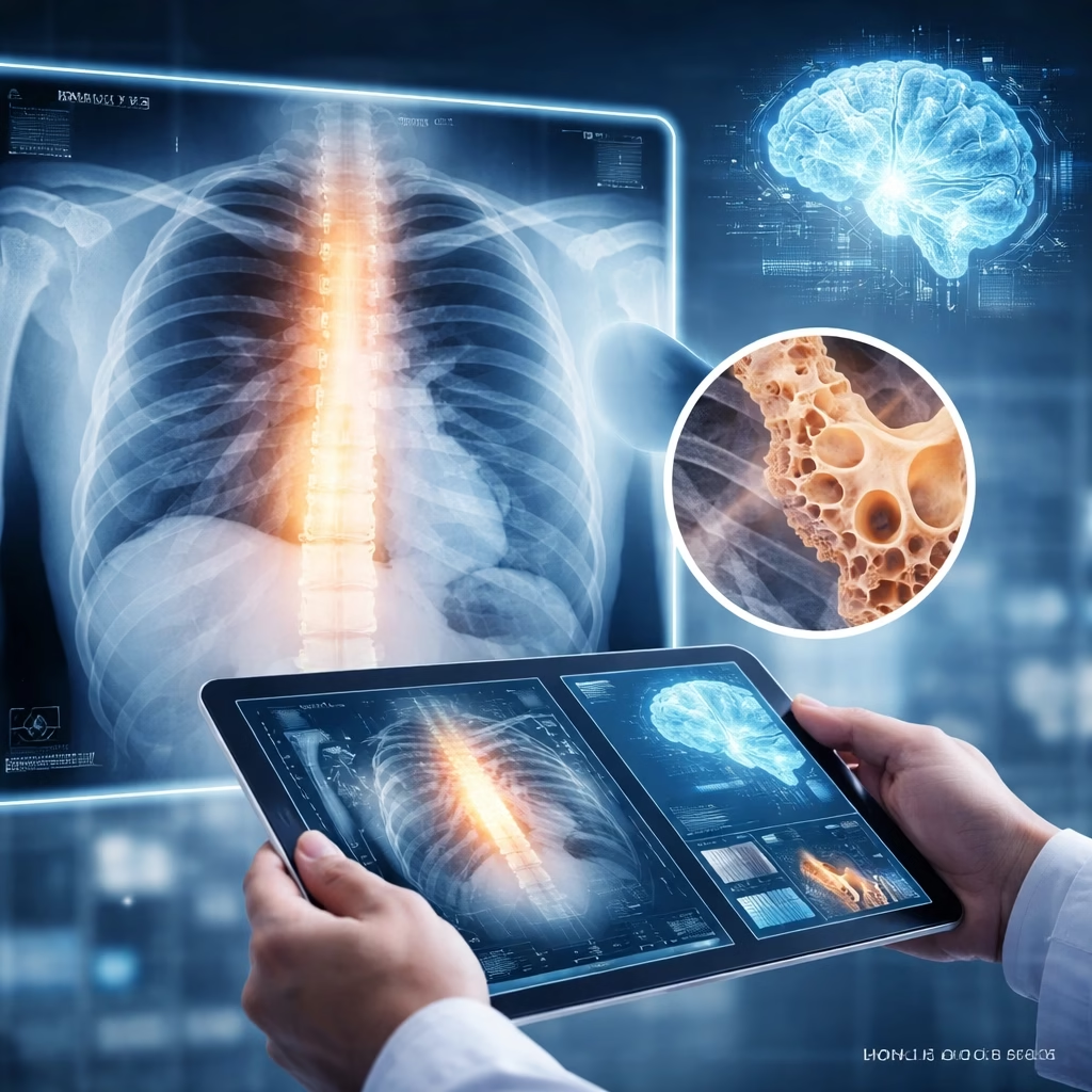
Key Developments: Knowledge Distillation to the Rescue
The geneonline.com team tackled this challenge using knowledge distillation, a technique where a large, complex "teacher" model transfers its knowledge to a smaller, more efficient "student" model. The teacher model was trained on a large, diverse dataset of chest X-rays, including images with varying degrees of bone density. This teacher model learns to identify subtle patterns indicative of osteoporosis.

The student model, significantly smaller and computationally less demanding, is then trained to mimic the teacher's predictions. This process allows the student model to achieve comparable accuracy to the teacher, but with reduced computational requirements. The team specifically focused on refining the student model to accurately identify areas of reduced bone density within the chest X-ray images.
Their research, published on the geneonline.com website, demonstrates that the knowledge distillation approach significantly improves the student model's performance compared to models trained directly on the chest X-ray data. The student model achieved an accuracy rate of 85% in detecting low bone mass, a notable improvement over previous methods using this type of imaging.
Impact: Broad Implications for Patient Care
This advancement has the potential to significantly impact patient care by enabling earlier and more accessible detection of osteoporosis. Integrating this AI technique into routine chest X-ray screening could identify individuals at risk who might otherwise remain undiagnosed.
More Read

Early diagnosis allows for timely interventions, including lifestyle modifications like increased calcium and vitamin D intake, and pharmacological treatments to slow bone loss and reduce fracture risk. This could lead to a reduction in the incidence of osteoporosis-related fractures, a major cause of morbidity and mortality, particularly among older adults.
The technology could also be particularly beneficial in resource-limited settings where DEXA scans are not readily available. Chest X-rays are a more widely accessible and affordable imaging modality.
What Next: Towards Clinical Implementation
The geneonline.com team is currently working on further refining the AI model and conducting clinical validation studies to assess its performance in real-world settings. Future steps include testing the model on diverse patient populations and integrating it into existing clinical workflows.
Clinical Validation Studies
The next phase involves rigorous clinical trials, planned to begin in early 2024, to evaluate the AI’s accuracy, sensitivity, and specificity in a larger patient cohort. These studies will also assess the impact of the AI-assisted diagnosis on patient outcomes.
Regulatory Approval
Following successful clinical validation, the team aims to seek regulatory approval from relevant health authorities like the FDA (Food and Drug Administration) for potential commercialization. They anticipate this process could take 1-2 years.

Expanding Applications
Beyond osteoporosis detection, the researchers believe that this knowledge distillation approach could be applied to other medical imaging modalities to improve diagnostic accuracy and efficiency. They are exploring potential applications in detecting other bone diseases and conditions affecting the chest, such as pneumothorax.


